Background
The presence of positively and negatively charged amino acids, as well as negatively charged glycans (sialic acids), indicates that large proteins exist as numerous charged species, and there are several side reactions that might cause a change in net charge. It is critical to understand which amino acids or glycans are involved and their specific location within a large biotherapeutic protein. Variants within an antibody's antigen binding region are more likely to have a significant impact on function. Ion exchange chromatography can separate some charge variations, particularly those found on the protein's surface (rather than hidden within the structure).
Nonetheless, distinguishing between a molecule with a net charge of +50 and a variation with a charge of +49 or +51 remains significant. Eliminating pore structure and thus slower diffusion kinetics by using nonporous particles, helps to improve peak shape and resolution. As a general approach, weak cation exchange columns are frequently used in charged variant analysis, alongside extensive method tuning.
Getting Started
As most proteins contain more basic amino acids than acidic amino acids, most charge variant separations necessitate cation exchange. However, each protein is unique, and determining the conditions that will provide the required resolution, will certainly necessitate significant optimisation. Strong cation exchange columns are often easier to work with, due to their permanent charge which is not influenced by eluent pH. However, for monoclonal antibodies, a weak cation exchange column may be the only option to attain the requisite resolution, through the manipulation of eluent pH and therefore the extent of stationary phase charge.
Before starting method development, it is critical to establish the target protein's isoelectric point, or pI. If the initial mobile phase pH is too close to the protein's pI, it will not be retained by the stationary phase. Depending on the magnitude of variation in pI between the variants of interest, the eluent pH may need to be 0.5 to 2 pH units away from the primary species' isoelectric point. Proteins can be eluted using a salt gradient (high ionic strength to disrupt protein adsorption to the column) or a pH gradient (proteins elute when the eluent pH is equal to the pI).
It is worthwhile investigating multiple columns during method development, as it is difficult to predict the consequence of even minor changes to method conditions, such as ionic strength and pH; both will affect the net charge on the protein and, in the case of weak ion exchange columns, the net charge on the column. A rigorous "Quality by Design" approach is recommended, and the use of experimental design approaches can be beneficial. Buffer advisor software can significantly reduce method development time, through online formation and manipulation of eluents of different ionic strength and composition.
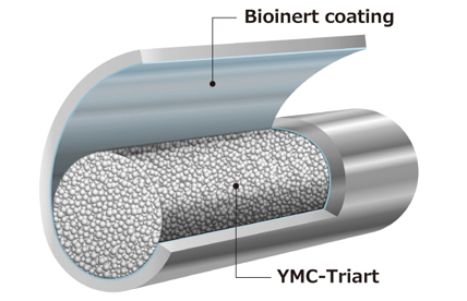
When the optimal separation conditions necessitate very low ionic strength buffers at pH levels at the extreme boundaries of the buffering range, PEEK columns may also be recommended.
Ion exchange chromatography, like size exclusion chromatography, is normally nondenaturing; the separation takes place on the intact, native protein.
Introduction
Proteins are composed of chains of amino acids, many of which have acidic or basic side chain functions. This produces an overall charge on the protein's surface, which may be adjusted by altering the pH of the surrounding solution. The isoelectric point (pI) is the pH at which the protein's net charge is neutral (number of positive charges equals number of negative charges). If the pH is less than this number, the protein has an overall positive charge and can be retained on a negatively charged cation-exchange sorbent; if the pH exceeds the pI, the protein has an overall negative charge and can be retained on an anion-exchange sorbent.
This paper discusses ion-exchange (IEX) chromatography, column selection options, critical mobile phase concerns, general IEX usage guidelines, instrument considerations, and more.


Figure 1 shows the effect of pH on net protein charge (Courtesy of YMC Europe, GmBH, Germany)
Separation based on ionic charge is normally conducted under nondenaturing conditions.
Ion-exchange is a popular approach for separating biomolecules depending on the extent of electrostatic charge. It is a gentle, non-denaturing approach that does not require organic solvents and is thus widely used for protein characterisation in their native or active state, as well as purification.
Proteins possess a range of functionalities, which can cause differences in charge. Acidic groups include C-terminal carboxylic acids, acidic side chains of aspartic and glutamic acid, and acidic groups derived from sialic acid in glycosylated proteins, while basic groups include N-terminal amines and basic side chains of arginine, lysine, and histidine. The overall charge of the molecule is thus determined by the pH of the surrounding solution, which influences the ion-exchange mechanism that can be used.


Figure 2 shows the separation mechanism of ion-exchange. The proteins are eluted by increasing the ionic strength (salt concentration) of the buffer to increase the competition between the buffer ions and proteins for charged groups on the IEX resin (courtesy of Agilent, United States)
The ion-exchange approach is thus appropriate for separating proteins with different isoelectric points, but it is also useful in differentiating charged isoforms of the same protein. In the increasingly significant field of biopharmaceuticals, where proteins are bioengineered and separated from fermentation reactions, it is critical to detect charged isoforms, which signal a variation in the protein's primary structure. A change in primary structure could signal a change in glycosylation or degradation pathway shift, such as loss of C-terminal residues or amidation/deamidation. They can also cause a shift in stability or activity, which could lead to immunologically adverse reactions. Ion exchange is utilised to isolate and quantify charge variations throughout the development process, as well as for quality control and assurance during biotherapeutic manufacturing. With large molecules like monoclonal antibodies (mAbs), it is also important to consider the molecule's size and structure (mAbs are generally 150 kD), especially since chromatographic interactions only occur with surface charges.


Figure 3 shows that charged variants of monoclonal antibodies arise through different levels of glycosylation, deamidation, and oxidation of amino acids, and through lysine truncation of heavy chains (courtesy of Agilent, United States)
Understanding the conditions for a successful ion-exchange separation.
Step 1:
Sample preparation
Sample preparation for ion-exchange chromatography is similar to that for other protein analyses. The sample must be soluble in the eluent and, ideally, dissolved in the mobile phase itself to prevent peak shape deformations. To protect the column from potential damage, it is recommended that samples be filtered before use to eliminate particulates. However, filtration should not be used to compensate for poor sample solubility; an alternative eluent may be required.
Low Protein Binding Filters
Low protein binding filters, such as the Agilent Captiva Premium PES Syringe Filters, offer exceptional and consistent low protein binding for protein filtration. For the majority of HPLC sample preparation, polyethersulfone (PES) filter membranes outperform polyvinylidene difluoride (PVDF) membranes. PES filters are compatible with standard LC solvents in the same way that PVDF filters are, but they are cleaner and have improved low binding performance.


Figure 4 shows Agilent Captiva Premium PES Syringe Filters (courtesy of Agilent, United States)
Step 2:
Column selection - Ion-Exchange
Column Media Choice
As with most chromatographic procedures, there are several columns to choose from. The initial concern in ion exchange should be "anion or cation-exchange?" There is also a choice between strong and weak ion exchange. In most cases, it is advisable to begin with a strong ion exchange column. Weak ion exchangers can then be utilised to provide a selectivity difference as needed.
The functional group in a strong cation-exchange column is sulfonic acid, therefore the stationary phase is negatively charged in all but the most acidic mobile phases. In contrast, the functional group in a strong anion-exchange column is a quaternary amine group, which is positively charged except in the most basic mobile phases. Strong ion-exchange columns offer the broadest operating pH range. Weak ion-exchange sorbents (carboxylic acids in weak cation exchangers and amines in weak anion exchangers) are more sensitive to the mobile phase conditions. The functions are analogous to the charged groups on proteins, and the degree of charge is regulated by both ionic strength and mobile phase pH.
This can result in a shift in resolution, which can be subtly controlled and optimized by carefully selecting operating conditions. Weak ion exchangers are thus an extra tool that can provide selectivity that a strong ion exchange column cannot.


|
Column
|
|
|
Eluent
|
A) 20 mM MES-NaOH (pH 5.6)
B) 20 mM MES-NaOH (pH 5.6) ) containing 05M NaCl
|
|
Initial Conc.
|
35 % B (70 mM NaCl)
|
| Gradient |
0.25 % B/min (0.5 mM NaCl)
|
|
Flow Rate
|
180 cm/hr
|
|
Temperature
|
30°C
|
|
Detection
|
UV at 280 nm
|
|
Injection
|
10 µL
|
|
Sample
|
Humanised monoclonal
IgG1 (1 mg/mL)
|
Figure 5 shows the analysis of a monoclonal antibody IgG1 using a strong cation exchanger (YMC BioPro IEX SF) and a weak cation exchanger. It demonstrates that the use of the SCX column results in a clearly higher resolution and more charge variants can be separated from the main peak. Furthermore, the run time can be reduced by nearly 50 % in combination with the smaller particle size and a shorter column length. (Courtesy of YMC Europe GmBH, Germany)
Pore Size
Porous materials have excellent binding capacity, high efficiency, and low operating pressure. Some fully porous stationary phases have large pores (1000 or 4000 Å). It is critical that the pores are large enough to allow proteins to permeate the structure completely. This results in a larger surface area and hence a higher loading capacity, making it better suited for preparative separations.
Nonporous particles provide greater resolution by eliminating rate limiting diffusion within the pore structure, thus producing narrower (more efficient) peaks. Smaller particle sizes have a larger charged surface area, which allows for a better binding capacity. Coupling nonporous particles with lower particle sizes and decreasing column lengths significantly reduces runtimes while increasing throughput.


| Columns | |
| Eluent | A) 20 mM NaH2PO4-Na2HPO4 (pH 6.8) B) 20 mM NaH2PO4-Na2HPO4 (pH 6.8) containing 0.5M NaCl |
| Gradient | 0 – 100 % B (0 – 60 min) |
| Flow Rate | 0.5 mL/min |
| Temperature | 25°C |
| Detection |
UV at 280 nm |
| Injection | 100 µL |
| Sample | Lysozyme |
Figure 6 illustrates how non-porous columns offer outstanding column efficiency for small sample loading amounts. These columns are especially suitable for microscale analysis, which requires higher resolution. Porous columns maintain good peak shape even when the loading amount increases. These high-capacity columns are useful for high-load analytical separations and laboratory-scale purification. (courtesy of YMC Europe, GmBH, Germany)
Particle Size
Particle size is an essential factor in column selection. Smaller particle sizes enable more efficient separation, albeit at the expense of increased operating pressure. Biomolecules are relatively large and have slower diffusion rates, so smaller particle sizes may not provide the same amount of resolution improvement as is seen with small molecules. Furthermore, eluents containing aqueous buffers are relatively viscous, therefore care must be taken to avoid high back pressures.


| Column A | Bio WCX, stainless steel 5190-2445, 4.6 x 250 mm, 5 µm |
| Column B | Bio WCX, stainless steel 5190-2443, 4.6 x 50 mm, 3 µm |
| Eluent |
A) 20 mM sodium phosphate (pH 6.5)
B) A + 1.6 M NaCl Gradient: 0 min – 100 % A : 0 % B |
| Gradient | 0 – 50 % B |
| Temperature |
Ambient |
| Injection |
10 µL |
| Detection |
UV at 220 nm |
| Sample | 0.5 mg/mL |
Figure 7 shows protein separation on Agilent Bio WCX columns (4.6 x 50 mm, 3 µm and 4.6 x 250 mm, 5 µm) at a flow rate of 1 mL/min. The separations are equivalent, but faster analysis times were achieved with smaller particle size and shorter column length – samples eluted from the longer column in 17 minutes but only in 12 minutes from the shorter column (courtesy of Agilent, United States)
Column Hardware
Stainless steel columns are commonly utilised, although salt gradients can be aggressive and induce corrosion if left in contact with the column. PEEK columns do not suffer from this issue and can be useful for metal-sensitive compounds, albeit operating at lower back pressures. A metal-free sample flow path can be achieved using a PEEK column and a bio-compatible instrument.
Column Diameter
The column diameter can also be essential, depending on the amount of sample being tested. If only a small amount of material is available, 2.1 mm id columns (at 0.35 mL/min) are suitable. However, when utilising smaller id columns, system volumes between the column and detector should be kept to a minimum to avoid severe dispersion and resolution loss.
Element Lab Solutions offer a wide range of IEX phases, internal diameters, particle sizes and hardware dimensions, enabling end users to select the optimum chemistry and hardware for a successful separation, regardless of the application type. Element technical specialists are on hand to guide you through the options and aid in the optimum selection for the application.
| Manufacturer | Description | Phase | Particle Type | Particle Size | Pore Size | pH Range | Max Temperature |
| Agilent | Bio IEX |
SCX
WCX
SAX
WAX
|
Non- Porous | 1.7, 3, 5 and 10 µm | N/A | 2 - 12 | 80°C |
| Bio mAb | WCX | Non- Porous | 1.7, 3, 5 and 10 µm | N/A | 2 - 12 | 80°C | |
| Bio-Monolith QA | SAX | Monolith | N/A | N/A | 2 - 11 | 40°C | |
| Bio-Monolith DEAE | WAX | Monolith | N/A | N/A | 2 - 11 | 40°C | |
| Bio-Monolith SO3 | SCX | Monolith | N/A | N/A | 2 - 11 | 40°C | |
| PL-SAX | SAX | Fully Porous PS/DVB | 5, 8, 10and 30 µm | 1000Å and 4000Å | 1 – 14 | 80°C | |
| PL-SCX | SCX | Fully Porous PS/DVB | 5, 8, 10and 30 µm | 1000Å | 1 – 14 | 80°C | |
| Zorbax SAX | SAX | Fully Porous | 5 µm | 70Å | 2 - 7 | 80°C | |
| Zorbax 300 SCX | SCX | Fully Porous | 5 µm | 300Å | 2 - 7 | 80°C | |
| Thermo Scientific | MAbPacSCX-10 | SCX | Non-porous | 5 and 10 µm | N/A | 2 - 12 | 60°C |
| MAbPacSCX-10RS | SCX | Non-porous | 5 µm | N/A | 2 - 12 | 60°C | |
| ProPac Elite WCX | WCX | Non-porous | 5 µm | N/A | 2 - 12 | 60°C | |
| YMC | BioPro IEX QA | SAX | Porous | 5 µm | 1000Å | 2 - 12 | 60°C |
| BioPro IEX SP | SCX | Porous | 5 µm | 1000Å | 2 - 12 | 60°C | |
| BioPro IEX QF | SAX | Non-porous | 5 µm | N/A | 2 - 12 | 60°C | |
| BioProIEX SF | SCX | Non-porous | 5 µm | N/A | 2 - 12 | 60°C | |
| Shodex | IEC QA-825 | SAX | Porous | 12 µm | 5000Å | 2 - 12 | 50°C |
| IEC DEAE-825 | WAX | Porous | 8 µm | 5000Å | 2 - 12 | 50°C | |
| Ashipak ES-502N 7C | WAX | Porous | 9 µm | 2000Å | 2 - 12 | 50°C | |
| IEC SP825 | SCX | Porous | 8 µm | 5000Å | 2 - 12 | 50°C | |
| IEC SP-FT 4A | SCX | Non-porous | 2.7 µm | N/A | 2 - 12 | 45°C | |
| IEC CM-825 | WCX | Porous | 8 µm | 5000Å | 2 - 12 | 50°C | |
| Ashipak ES-502C 7C | WCX | Porous | 9 µm | 2000Å | 2 - 12 | 50°C | |
| CXpak P-421S | SCX | Non-porous | 6 µm | N/A | ≥3 | 63°C |
Table 1 shows the range of analytical IEX columns available from Element Laboratory Solutions (Courtesy of Element Laboratory Solutions, Strathaven, UK)
Step 3:
HPLC system considerations
Charged variant analysis is best performed with bio-inert/compatible LC equipment, such as the Agilent 1260 Infinity Bio-inert Quaternary LC or the ThermoFisher Vanquish Flex UHPLC. These LC systems can easily manage challenging solvent conditions, such as pH values ranging from 1 to 13, as well as buffers with high salt concentrations. Corrosion-resistant materials, such as titanium and MP35N alloy, are employed in the solvent delivery system, while metal-free components in the sample flow path result in very low or no protein binding.


Figure 8: Agilent 1260 Infinity Bio- Inert Quaternary LC
(courtesy of Agilent, United States)


Figure 9: Thermo Scientific Vanquish Biocompatible Flex UHPLC
(courtesy of Thermo Scientific, United States)
Detection
IEX-UV
UV detection at 210 nm or 220 nm provides the maximum signal intensity and sensitivity for many proteins, which are made up of numerous amino acids connected together by amide bonds. However, some of the eluents commonly used in ion exchange have a high background absorbance at low wavelengths, thus 254 nm or 280 nm may be used instead. These wavelengths are only sensitive to amino acids that have aromatic or highly conjugated side chains, resulting in significantly lower sensitivity.


| Column A | Bio SCX, stainless steel 5190-2423, 4.6 x 50 mm, 3 µm |
| Column B | Bio WCX, stainless steel 5190-2443, 4.6 x 50 mm, 3 µm |
| Column C | Bio mAb, stainless steel 5190-2403, 4.6 x 50 mm, 3 µm |
| Eluent | A) 10 mM Sodium phosphate, pH 5.7 B) A + 1 M NaCl |
| Flow Rate | 0.5 mL/min |
| Gradient | 0 – 100% B (0 – 25 min) |
| Detection | UV at 254 nm |
| Injection | 1 µL (0.25 mg/mL) |
| Temperature | Ambient |
| Sample | Ribonuclease A Cytochrome C Lysozyme Protein mix |
Figure 10 shows the separation of protein standard on Agilent 3 µm ion-exchange columns by cation-exchange chromatography (Courtesy of Agilent, United States)
CX-MS
Native protein analysis using cation-exchange chromatography coupled to mass spectrometry (CX-MS) is a reliable method for identifying microheterogeneities in biopharmaceuticals, notably monoclonal antibodies. CX-MS enables charge-variant analysis to detect chemical and enzymatic changes that occur during mAb synthesis, purification, formulation, and storage. These alterations can impact the structure and biological action of medicines, thus they must be characterised properly. Previously, ion-exchange chromatography (IEX) was incompatible with mass spectrometry due to the use of non-volatile salts with high ionic strength. However, the introduction and improvement of volatile, salt-mediated pH gradients creates an opportunity for CX-MS.


| Column A | BioPro IEX SF, SF00S05-1046WP, 100 x 4.6 mm, 5 µm |
| Eluent | A) 20 mM ammonium acetate, pH 5.6 B) 140 mM ammonium acetate, 10 mM ammonium carbonate, pH 7.6 |
| Flow Rate | 0.4 mL/min |
| Gradient | 0 % B (0 – 2 min), 0 – 100 %B (2 – 16 min), 100 % B (16 – 20 min), 100 % A (20 – 27 min) |
| Temperature | 45 °C |
| Equipment | Post column analytical splitter ( ~400:1) to reduce analytical flow to ca. 1 µL/min |
| Detector | PDA (UV), PicoTip Emitter (NSI-MS) |
Figure 11 shows TICs of the SCX-MS analysis of 6 different mAbs with pI values ranging from 6.3 to 9.2. The combination of IEX and MS necessitates a unique configuration in which a stainless-steel T-piece following the column separates the flow. Most of the flow is routed to the UV detector, with the residual sub-microlitre per minute flow going to the nanoelectrospray ionization mass spectrometer (NSI-MS). NSI is employed because it can withstand salt concentrations as high as 600 mM ammonium acetate. Isopropanol is employed as a dopant, or modified desolvation gas, to improve spray stability even more (courtesy of YMC Europe, GmBH, Germany)
AEX-MS
Cation exchange chromatography (CEX) is a great tool for determining charge heterogeneity in biomolecules. This also holds true for most commercially available monoclonal antibodies (mAbs). They are often IgG1-based and have a high isoelectric point (pI) of ≥8. As a result, CEX combined with mass spectrometry (MS) is the traditional method. In contrast, anion exchange chromatography (AEX) has only been employed for proteins that are somewhat acidic. However, for IgG4-based mAbs, AEX could be an alternative. They have a pI < 8, making CEX less acceptable.


Figure 12 shows the schematic setup of the developed native AEX-method (courtesy of YMC Europe, GmBH, Germany)


| Column | BioPro IEX QF, QF00S05-1046WP, 4.6 x 100 mm, 5 µm |
| Eluent | A) 10 mM ammonium acetate, pH 6.7 B) 300 mM ammonium acetate, pH 6.8 |
| Gradient | 0 % B (0 – 2 min), 0 – 100 % B (2 – 18 min), 100 % B (18 – 22 min) |
| Temperature | 45°C intact mAb, 25°C subunit analysis |
| Detection | NSI-MS (nanoelectrospray ionization) |
| Sample | In-house IgG4 based mAbs (Regeneron) NIST mAb |
| Post Column Setup | Post column stainless-steel T-piece to direct the majority to the UV detector. Remaining sub-microlute per minute flow directed to the NSI-MS |
| Flow Rate | 0.4 mL/mi |
| Injection | 5 or 10 µg mAb sample |
Figure 13 shows the conditions that were applied to various IgG4-based mAbs (pI 6.1–7.3) as well as to the NISTmAb, which is IgG1-based and has a pI of 9.2. The figure shows that the AEX-MS method is suitable for IgG4-based mAbs with moderate pIs, but not for IgG1-based mAbs with higher pIs. The separation improves as the pI gets lower. mAb-6 has a pI of 7.3, which is higher than the mobile phase pH, but sufficient separation still occurs. This suggests that it is the surface charge rather than the intrinsic charge that causes the AEX-based separation (courtesy of YMC Europe, GmBH, Germany)
Step 4:
Flow Rate
The typical flow rate for 4.6 mm id columns is 0.5 to 1.0 mL/min. For some applications, speed of analysis is critical. Shorter columns (50 mm instead of 150 mm or 250 mm) can be used to minimize analysis time, as can higher flow rates (while not exceeding column pressure constraints).
The performance of a column, as measured by plate count, is dependent on particle size and column length. From this it may be inferred that a shorter column packed with smaller particles can be used to achieve the same level of performance when compared to a longer column packed with larger particles. This is commonly found in practice. However, for gradient elution, further modifications to the method need to be employed to provide the additional benefits of shorter run times and greater productivity.
Converting gradient times into column volumes is a useful way of calculating the shorter gradient program and can provide the desired outcome in terms of higher speed separations. However, smaller particle sizes may require higher flow rates to attain maximum performance, as described by van Deemter curves. Increasing the flow rate should mean that it is possible to further reduce the gradient time.


| Column A | Bio WCX, stainless steel 5190-2443, 4.6 x 50 mm, 3 µm |
| Eluent | A) 20 mM sodium phosphate (pH 6.5) B) A + 1.6 M NaCl |
| Gradient | 0 – 50 % B (scaled according to flow rate) |
| Sample | Ovalbumin (1), Ribonuclease A (2), Cytochrome c (3), Lysozyme (4) |
| Temperature | Ambient |
| Injection | 10 µL |
| Detection | UV at 220 nm |
| Flow Rates | 1.0, 1.5, 2.0, 2.5 mL/min |
Figure 14 shows a 4-minute gradient separation (gradient was scaled by converting gradient times into column volumes) was carried out at 1.0, 1.5, 2.0 and 2.5 mL/min. As expected, the higher liner velocity created from higher flow rates improved the peak shape (courtesy of Agilent, United States)


| Column A | Bio WCX, stainless steel 5190-2443, 4.6 x 50 mm, 3 µm |
| Eluent | A) 20 mM sodium phosphate (pH 6.5) B) A + 1.6 M NaCl |
| Gradient |
0 – 50 % B (scaled according to flow rate) |
| Sample | Ovalbumin (1), Ribonuclease A (2), Cytochrome c (3), Lysozyme (4) |
| Temperature | Ambient |
| Injection | 10 µL |
| Detection | UV at 220 nm |
| Flow Rates | 1.7 mL/min |
Figure 15 shows how utilising smaller particle size (1.7 µm) can lead to significant reductions in run times, from 20 or 30 minutes down to less than 3 minutes. It was found that at a flow rate of 1.7 mL/min, the backpressure remained below 400 bar and still provided exceptional peak shape and resolution (courtesy of Agilent, United States)
Step 5:
Mobile Phase Selection
The initial mobile phase selected, will be determined by the protein's pI and the technique of analysis, i.e. cation- or anion-exchange. The buffer's job is to limit the pH variation during separation, ensuring that the compounds being examined have a consistent charge. It is critical to remember that a buffer will only fulfill this function if it is within one pH unit of its dissociation constant, pKa. Phosphoric acid or phosphates have three dissociation constants:


Figure 16 shows the three dissociation constants of phosphoric acid or phosphates (courtesy of Agilent, United States)
Phosphate buffers with a pH of 6 to 7 are ideal for cation-exchange chromatography, typically at concentrations of 20 to 30 mM, and have a low background absorbance at 210 nm. It is critical to prepare buffers in a systematic and accurate manner, as minor changes in ionic strength or pH can alter protein retention time to varying degrees, resulting in changes in selectivity and non-robust chromatographic resolution.
Unlike strong ion-exchange columns, which are fully ionized under normal operating conditions, weak ion-exchange columns can be ionized to varying degrees depending on buffer pH and ionic strength. This is one of the options available to change selectivity and achieve the necessary separation.
To elute biomolecules from the column, a competitive ion must be added. This is typically performed using a linear salt gradient. Eluent A will include the buffer that has been adjusted to the right pH. Eluent B will include the same buffer concentration but with a higher concentration of sodium chloride, say 0.5 M, and the pH will be adjusted to the same value.
Gradient Selection
Salt Gradient
In salt gradient IEX, the buffer's pH is fixed. In addition to selecting the optimum pH for the buffer, the initial ionic strength is kept low since proteins' affinity for IEX resins decreases as ionic strength increases.
Proteins are eluted by raising the buffer's ionic strength (salt concentration), which increases competition between buffer ions and proteins for charged groups on the IEX resin. As a result, the interaction between the IEX resin and the proteins is diminished, causing the proteins to dissociate from the stationary phase surface.


| Column | Propac Elite WCX, 4.0 x 150 mm, 5 µm |
| Eluent | A) 20 mM MES, pH 6.5 B) 20 mM MES + 500 mM NaCl, pH 6.5 |
| Flow Rate | 1.0 mL/min |
| Gradient | 0 min Loading, 0.8 min Loading, 1.0 min Initial, 16.0 min Final, 16.1 min 50 % B, 18.0 min 50 % B, 18.1 min Loading, 25.0 min Loading |
| Temperature | 30°C |
| Detection | UV at 280 nm |
| Injection | 3 µL |
| Sample | See Table X |
Figure 17 shows the analysis of innovator mAbs and respective biosimilars using general salt gradient (courtesy of Thermo Scientific, United States)
|
mAb
|
Loading %
|
Initial %
|
Final %
|
ΔB/Δt %/min
|
Minutes Reduced |
| Rituximab Rituximab biosimilar |
8 | 13 | 22 | 0.60 | 8.3 |
| Trastuzumab Trastuzamab biosimilar |
9 | 12 | 20 | 0.53 | 5.7 |
| Bevacizumab Bevacizumab biosimilar |
8 | 10 | 17 | 0.47 | 4.3 |
| Vedolizumab | 5 | 7 | 14 | 0.47 | 4.3 |
| Infliximab | 4 | 8 | 18 | 0.67 | 6.0 |
| Secukinumab | 5 | 10 | 16 | 0.40 | 12.5 |
| Pertuzumab | 9 | 12 | 20 | 0.53 | 5.7 |
Table 2: %B gradient values for loading, initial and final; gradient slope and time (minutes) reduced with step change (courtesy of Thermo Scientific, United States)
pH Gradient
In pH-gradient-based IEX, the starting buffer is kept at a constant pH to guarantee that the proteins have an opposing charge to the stationary phase and so bind to the column. The pH of the buffer changes over the gradient, causing the proteins to transition to a net zero charge (pI) and eventually to the same charge as the resin, allowing the protein to be released and eluted from the column.
In the fast-paced drug development setting, a platform approach that can accommodate the bulk of mAb analyses is desired. Significant method development is necessary to optimize the salt gradient for charge variant separation of each mAb. Ion exchange separations by pH gradient have the benefit of a generic platform approach, which saves time on method development. Additionally, heavy salts are removed from LC systems, resulting in reduced cleaning and downtime.
One of the difficulties in pH-gradient separations is selecting a buffer solution that can cover a large pH range while producing a linear pH gradient. Many vendors, including Agilent, Thermo, and YMC, now provide innovative pH gradient applications and buffer solutions. Once the approximate pH elution range of the target mAb has been determined in a screening experiment, the separation is optimized by running a shallower pH gradient over a smaller pH range.


| Column | ProPac Elite WCX, 4.0 x 150 mm, 5 µm |
| Eluent | A) CX-1 pH gradient buffer A (pH 5.6) B) CX-1 pH gradient buffer B (pH 10.2) |
| Gradient | 20 – 70 % B (0 -15 min), 70 – 100 % B (15 – 15.1 min), 100 % B (15.1 – 17 min), 100 – 20 % B (17 – 17.1 min), 20 % B (17.1 – 204 min) |
| Sample | Rutiximab, Trastuzumab, Adalimumab biosimilar, Bevacizumab, Infliximab, Secukinumab, Pertuzumab, Vedolizumab |
| Temperature | 30°C |
| Detection | UV at 280 nm |
| Injection | 2 µL (5 mg/mL) |
| Flow Rate | 0.8 mL/min |
Figure 18 shows the separation of IgG1 therapeutic mAbs using a 20 – 70% B pH gradient. The combination of the Thermo Scientific™ ProPac™ Elite WCX column and Thermo Scientific™ CX-1 pH Gradient Buffers provides a high-resolution, fast, easy-to-optimize, and reproducible platform method for charge variant characterization of therapeutic mAbs. (courtesy of Thermo Scientific, United States)
Summary: Developing an Effective Ion-Exchange Method
It should be remembered that biomolecules such as monoclonal antibodies are extremely complicated. A typical mAb contains more than 1,300 unique amino acids. Approximately 130 have acidic side chains, while 180 have basic residues. A monoclonal antibody is likely to have a net positive charge at neutral pH, hence it should be separated with a cation-exchange column. However, it is difficult to determine the real isoelectric point, pI, of such a molecule, hence some method development or optimization is required.
Sample Preparation
- Ideally, samples should dissolve in the mobile phase (eluent A).
- If the sample is cloudy, adjust the mobile phase conditions.
- Filtration and centrifugation can clarify samples but may alter their composition.
- Analyse freshly prepared samples as soon as possible. Refrigeration can extend the "shelf life" of samples.
- Bacterial growth can develop quickly in buffer solutions.
Column Media Choice
- The isoelectric point of the protein(s) of interest determines whether to use anion or cation exchange.
- Strong ion-exchangers are the preferred choice, but weak ion-exchangers can provide additional selectivity if necessary.
Column Selection
- Proteins of interest should be able to freely permeate porous particles. Non-porous spherical particles provide the best resolution for analytical separations where column loading capacity is not a major consideration.
- Particle size: use smaller particles for higher resolution (which results in higher back pressure).
- Column length: Use shorter 50 mm columns for faster separation with smaller particles, or longer 250 mm columns for higher resolution.
- Use smaller ID columns to reduce solvent consumption and injection volume, especially if sample size is limited.
Mobile Phase
- To maintain the correct operating pH, the mobile phase should include buffer (usually 20 mM). The pH and ionic strength of the buffer can influence resolution on weak ion-exchange products, so the best conditions should be determined experimentally.
- Adding sodium chloride to the mobile phase will change the pH. Re-adjust as necessary.
- Use new mobile phase quickly as dilute buffer at room temperature promotes rapid bacterial growth.
- Buffer shelf life is less than seven days unless refrigerated.
- Filter before use. Particulates can be present in water (less likely) or in buffer salts (more likely).
Column Conditioning and Equilibration
For reproducible ion-exchange separation, the column equilibration and gradient cleanup phases are crucial. Protein elution is accomplished by increasing ionic strength, changing eluent pH, or both, therefore the column must be equilibrated at the end of each analysis to the starting conditions, ionic strength, and pH. If this is not done, the next column used will have a different profile, as the protein will interact differently with the column.

